DESCRIPTION AND BACKGROUND
Symptoms of impietratura were first described by Reichert and Hellinger (1930) in Palestine. The disease was called "samrah" by the growers, and the affected trees produced fruit with gumming in the albedo of citron, grapefruit and oranges. Ruggieri (1955) named the disease "impietratura" because the fruit turned hard like a stone. For further description of the disease see Catara and Scaramuzzi (1980), Catara et al. (1977) and Papasolomontos (1969). Seasonal variation in appearance of symptoms has been noted, and strain differences reported.
The disease is prevalent in all countries of the Mediterranean basin and also in Iran, Venezuela, India and South Africa. There is no reason why the disease cannot exist anywhere citrus is grown if diseased budwood is introduced and propagated. The fruit of sweet orange, grapefruit and Volkamer lemon are highly susceptible. The disease has been found on fruit of lemon, rough lemon, bergamot, tangelo, citron and mandarin.
The pathogen is believed to be a virus but has not been isolated or characterized. It may be related to, or part of, the concave gum family that produces a distinct oak-leaf pattern (OLP) on leaves in the spring flush of growth on field trees or leaves of seedling indicators. The OLP symptom in inoculated seedlings was shown to be diagnostic for impietratura by Bar-Joseph and Loebenstein (1970). The absence of OLP symptoms in indicator plants inoculated with tissue from heat-treated or shoot-tip grafted plants should indicate elimination of the pathogen from those plants. Thus, a short-term index using seedlings may be efficient for determining presence or absence of the disease.
Transmission of the disease is primarily by humans when propagating infected budwood. It is apparently not seed-transmissible (BarJoseph,1976), and there is no evidence of mechanical or vector transmission. The disease was transmitted by insertion of pollen from infected trees under the bark of Orlando tangelo test trees (Vogel and Bové,1980).
METHODS OF DETECTION Method 1: Field diagnosis
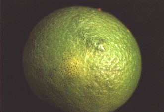
Certain symptoms associated with impietratura are highly diagnostic when observed in field trees. These are:
• The distinct discoloration of the rind, usually as
circular spots as shown in Figures 84 and 88, and the brown gum
spot found directly under the cut surface of the spot as shown in
Figures 85-87. These discoloured spots are usually seen as raised
bumps or protuberances on the rind.
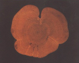
• If the fruit is sectioned, gum spots may appear throughout
the rind and albedo as shown in Figure 87.
• Appearance of many small fruit, which harden, drop to the
ground and, when cut, show gumming in the rind.
• The appearance of OLPs in the emerging leaves during the
first growth flush during the cooler spring months. This symptom,
in association with fruit symptoms, is helpful in diagnosis.
The appearance of all four of the above symptoms is diagnostic. Possible confusion with boron deficiency can be resolved by leaf analysis. A normal tree will contain 30-200 ppm boron in the dry matter of mature leaves, whereas a tree with boron deficiency will contain 3-25 ppm boron. In addition, the young leaves of boron deficient trees often show yellowish translucent spots, accompanied by marked leaf distortions or deformations, and old or mature leaves may show corky, split veins and midribs.
Method 2: Inoculation to field trees
The fruit symptoms associated with impietratura can be transmitted by grafting budwood from infected trees on to branches of field trees and the symptoms reproduced in the developing fruit.
Collection of budwood. Collect budwood, if feasible, from behind a fruit showing typical symptoms.
Inoculum tissue. "Buds" (buds, blind buds or chip buds).
Inoculation. Inoculate with ten to 15 "bud" grafts into a two- to three-year-old branch of a grapefruit tree which has fruited in the past. Any grapefruit tree, preferably on rough lemon or sour orange rootstock, is a superior indicator although other cultivars can be used (Papasolomontos and Economides,1967). Inoculate two to four branches on two trees in this manner and self-inoculate a third tree as a control.
Time of year. Graft inoculation should be carried out in the autumn, or as early in the spring as possible whenever the bark is slipping. If the bark is not slipping, chip buds can be used, preferably in February (see bottom bud in Figure 127 in Part II).
Inoculum survival. Cut the tapes covering the "buds" about six weeks after inoculation and record the "bud" survival. It is always good sanitary procedure to disinfect any tool used for cutting plant tissue in the laboratory or field by dipping it in a 1 percent sodium hypochlorite solution.
Temperature requirements. A cool spring followed by warm late spring and summer provides the best conditions for bringing out fruit symptoms (Bar-Joseph,1976). This is why late autumn or early spring inoculations are preferred.
Time for first symptoms. Bar-Joseph and Ben Shalom (1982) reported the development of symptoms on fruits in three to six months following inoculation. Symptoms should certainly appear by the next season. However, for possible mild-reacting isolates, observe for three full seasons in the field.
Method 3: Inoculation to indicator seedlings
Although OLP is generally diagnostic for the concave gum family of pathogens and is not specific for impietratura, the presence or absence of OLP in the young leaves of inoculated Dweet tangor, mandarin or sweet orange indicator seedlings may be diagnostic for the disease. This seedling index is useful for diagnosing the results of thermotherapy or shoot-tip grafting, since OLP is always associated with impietratura(Bar-Joseph and Loebenstein,1970). See Method 2 in the concave gum section for the detection of oak-leaf patterns (Figure 89) in seedling indicators
IMPIETRATURA DETECTION
Summary
Indicators:
Field: grapefruit/rough lemon or sour orange rootstocks for fruit
symptoms.
Seedlings: Dweet tangor, mandarin or sweet orange for OLP.
No. plants/test:
Field: minimum 2 trees; inoculate 4 branches per tree.
Seedlings: 4 Dweet tangor, mandarin or sweet orange (3 plus 1
control in each of 2 containers).
Inoculum:
"Buds" (buds, blind buds or chip buds).
Temperature: Field: cool spring followed by warm late
spring and summer.
Seedlings: 24-28°C max. day/18-21°C min. night.
Plant growth:
Seedlings: allow full flushes to develop after initial cutback.
First symptoms:
Field: 3 to 6 months in the developing fruit.
Seedlings: OLP in 5 to 8 weeks.
Symptoms:
Field: gum in fruit rind and albedo.
Seedlings: leaf fleck and OLP.
REFERENCES
Bar-Joseph, M.1976. Some effects of temperature on symptom appearance and therapy of citrus impietratura disease. In Proc. 7th Conf. IOCV, p. 105-108. Riverside, IOCV.
Bar-Joseph, M. & Ben-Shalom, J.1982. Limited systemic spread of impietratura and psorosis-A in graft-inoculated grapefruit trees. Plant Disease, 66: 820-821.
Bar-Joseph, M. & Loebenstein, G.1970. Leaf flecking on indicator seedlings with citrus in Israel. A possible indexing method. Plant Dis. Rep., 54: 643-646.
Catara, A. & Scaramuzzi, G.1980. Impietratura. In Bové,J.M. & Vogel, R.,eds. Description and illustration of virus and virus-like diseases of citrus. A collection of colour slides. Paris, I.R.F.A. SETCO-FRUITS.
Catara, A. et al.1977. Present status of impietratura disease. In Proc. Int. Soc. Citriculture, 3: 946-953.
Papasolomontos, A. 1969. A report on impietratura disease of citrus; its distribution and importance. Proc. 1st Int. Citrus Symp., p. 1457-1462.
Papasolomontos, A. & Economides, C.V.1967. Effect of rootstocks on the incidence of impietratura-diseased grapefruit fruits. Plant Dis. Rep., 51: 684-686.
Reichert, I. & Hellinger, E.1930. Internal decline. A new physiological disease of citrus fruits in Palestine. Hadar, 3: 220-224.
Ruggieri, G.1955. Le arance impietrate. Riv. Agrumic., 1(2): 65-69.
Vogel, R. & Bové, J.M.1980. Pollen transmission to citrus of the agent inducing cristacortis and psorosis young leaf symptoms. In Proc. 8th Conf. IOCV, p. 188190. Riverside, IOCV.
FIGURE 86 Gum showing in the albedo when the rind is sliced somewhat deeper
FIGURE 87 Impietratura-induced gumming in albedo of a sectioned young grapefruit (Spain)
DESCRIPTION AND BACKGROUND
Cristacortis was first described and named by Vogel and Bové (1964). Descriptions and illustrations of cristacortis are further documented by Vogel and Bové (1968,1980b).
Cristacortis is found primarily in the Mediterranean basin, i.e. Algeria, Corsica, Italy, Morocco, Sardinia, Spain, and probably in other parts of this region. Its presence elsewhere in the world is limited, but it could become established wherever citrus is grown if diseased budwood is imported. Susceptible varieties are sweet orange, mandarin, tangelos, tangors, grapefruit, sour orange, rough lemon, siamelo, sweet lime, and occasionally lemon. It has not been found in Troyer citrange, trifoliate orange, citron, chinotto, Citrus hystrix or Mexican lime.
The disease can be distinguished from cachexia by the type and variety of pits. In tangelo or mandarin, cristacortis pits are sharp, deep and distinct, with gum usually at the base of the pit (Figures 91, 92b and 93b). In contrast, the symptoms of cachexia disease in these varieties are undulating depressions and general gumming in the wood and bark (Cachexia, Figures 44 and 45). However, where both pathogens exist together in the same host, the individual diseases may be more difficult to diagnose except by indexing to specific sensitive indicator plants. When the outside trunk bark is observed, the indentations formed by the internal pits caused by cristacortis may resemble severe tristeza stem pitting. Again mixtures may exist, and indexing to specific indicators is needed to distinguish them. Cristacortis differs from concave gum by the quality of the pits, i.e. those of cristacortis being sharp and deep in contrast to the more rounded concavities and cupped deformations associated with concave gum (compare Concave gum, Figure 81 with Figures 90 and 92a).
The cristacortis pathogen has never been isolated but is presumed to be a virus. It may be related to the concave gum pathogen since both induce oak-leaf patterns (OLPs) in leaves of field trees or index plants. The OLP symptom is partially diagnostic and can be used for determining the presence or absence of the pathogen in a plant index text after heat treatment or shoot-tip grafting. Mild, moderate and severe forms of the disease exist and the Clementine mandarin is useful for strain identification(Vogel and Bové,1976).
The disease is transmitted primarily by humans via propagation of infected budwood or by topworking with budwood from infected trees. It is readily bud-transmitted to other citrus. Mechanical, vector and seed transmission have not been demonstrated.
Transmission has been accomplished by placing pollen from infected trees under the bark of indicator plants (Vogel and Bové,1980a).
METHODS OF DETECTION Method 1: Field diagnosis
The distinct pits in both the scion and the rootstock (Figure 90), coupled with the appearance of oak-leaf patterns in the young leaves of the field tree during the spring flush of growth (Figures 83 and 95), are highly diagnostic. However, confirmation should be made by indexing.
In the Mediterranean region, where cristacortis is prevalent, the sour orange is the predominant rootstock and will show the pitting symptoms of cristacortis (Figure 90). In contrast, cachexia-, concave gum- and impietratura-infected trees will not show these classical pitting symptoms in the sour orange rootstock. Therefore, the presence of these deep pits in sour orange rootstock is diagnostic for cristacortis.
Figure 91 shows a typical deep pit in the trunk wood with gum at the base of the pit, and its corresponding peg in the cut section of the bark of a Navelina orange tree in Spain.
Method 2: Indexing to tangelo as a budded scion
Orlando, Webber or Williams tangelo can be used as the scion and sour orange as the rootstock. The method of testing is similar to the forcing of a scion indicator bud described in the cachexia section. Buds taken from seedlings of known pathogen-free sources of these scion indicator varieties are propagated on sour orange rootstock, which is inoculated with "bud" grafts at the same time. The tangelo scion is then forced (Figure 47). Inoculated plants are held in a screenhouse under natural conditions (preferably cool). The detailed procedure is as follows:
Collection of budwood. Budwood to be indexed should be collected from four quadrants of a selected field tree. A minimum of four budsticks should be collected from each test tree.
Inoculum tissue. A minimum of two inoculum "buds" (preferably blind buds or bark pieces) should be grafted to the sour orange rootstock. The tangelo indicator bud to be forced is inserted about 25 cm above the soil surface and the two inoculum "buds" are grafted below the scion bud. The plant is then bent to force the tangelo bud (see procedure for forcing Parson's Special mandarin buds in the cachexia section and Figure 47).
Number of indicator plants. A minimum of four, but preferably six or eight, indicator plants should be used to test each candidate or selection. The inoculum "buds" cut from four budsticks collected around each tree should be evenly distributed among the four to eight indicator plants. After inoculation, inoculum-budwood should be retained and refrigerated for possible future use in the event of graft failure.
Controls. If available, mild- and severe-positive controls plus non-inoculated or self-inoculated negative controls should be included for each test or experiment. A minimum of four but preferably six plants for each of the controls should be used and possibly more for the mild-positive control.
Inoculum survival. Wrapping tapes should be removed two to three weeks after inoculation and the inoculum tissue observed for graft survival. Although cristacortis is not known to be mechanically transmissible, cutting tools should be routinely disinfected in 1 percent sodium hypochlorite solution between plants. Any dead or dying inoculum buds should be recorded and if both buds are found dead, the plants should be regrafted (or a new plant inoculated) using inoculum stored in the refrigerator.
Post-inoculation care. The tangelo budling should be trained as a single shoot and tied to a stake (Figure 48). Temperatures should be from cool to moderate.
Location. Plants should be well spaced. Vogel and Bové (1972) report that in Corsica symptoms develop best under natural outdoor conditions rather than in a greenhouse. Vogel (personal communication) prefers a screenhouse in order to enhance maximum rapid symptom development.
Time for development of symptoms. Symptoms in tangelo will begin to show in eight to ten months, and trees inoculated with mild strains may take from 20 to 24 months.
Symptoms. Symptoms will first appear on the outer bark as small depressions (Figure 92a). If the bark is cut to expose the wood underneath, deep pits can be observed (Figures 91, 92b, 93b and 94). Young trees in the screenhouse should be examined in the spring during the first flush of leaf growth for OLP symptoms (Figure 95), and these recorded. The presence of OLP can be indicative of other associated pathogens, i.e. concave gum or impietratura, and its presence is not necessarily diagnostic for cristacortis. However, when OLPs are found in association with deep pits in the scion and rootstock of the indicator plants, or with pitting in the tangelo seedling as shown in Figure 94, the disease may then be diagnosed as cristacortis. The presence of cachexia may confuse the diagnosis somewhat, but the symptomatic deep pits in both scion and rootstock or in the tangelo indicator are very characteristic and diagnostic for cristacortis and not for cachexia. Also, as shown in Figure 93, cristacortis has a very different type of symptom from that of concave gum (or cachexia) when observed in cross-section.
Termination. When 75 percent of the inoculated mild-positive control plants show definitive symptoms, the bark from all test plants can be completely peeled and the wood examined for evidence of pitting.
Method 3: Observation for oak-leaf patterns in indicator seedlings
The more rapid seedling index for detection of leaf symptoms of OLP should be used if elimination of the pathogen is the primary objective (see procedures for detecting OLP symptoms under concave gum).
CRISTACORTIS DETECTION
Summary
Indicators:
Screenhouse: Orlando, Webber or Williams tangelo as scions
grafted on sour orange rootstock.
Seedling index: Dweet tangor, King, Kara or Dancy mandarin or
sweet orange seedlings.
No. plants/test:
Screenhouse: 4 to 8 tangelos, 1 per container.
Seedling index: 4 (3 plus 1 control in each of 2 containers).
Inoculum:
"Buds" or bark pieces.
Plant growth:
Screenhouse: train as a single shoot or leader or as a small
tree.
Seedling index: allow development of full flushes of growth after
initial cut-back.
Temperature:
Screenhouse: relatively cool.
Seedling index: cool 24-27°C max. day/18-21 °C min. night.
First symptoms:
Screenhouse: in tangelos: Severe 8 to 10 months; Mild - 20 to 24
months.
Seedling index: OLP in 5-8 weeks.
Symptoms:
Screenhouse: distinct sharp and deep pits in tangelo scion and
sour orange stock.
Seedling index: leaf fleck, OLP.
REFERENCES
Vogel, R. & Bové, J.M.1964. Stem pitting sur bigaradier et sur oranger tarocco en Corse: une maladie à virus. Fruits,19:269-274.
Vogel, R. & Bové, J.M.1968. A virus disease inducing stem pitting on sour orange and other citrus species. In Proc . 4th Conf. IOCV, p. 221 228. Gainesville, Univ. Fla. Press.
Vogel, R. & Bové, J.M.1972. Relationship of cristacortis virus to other citrus viruses. In Proc. 5th Conf. IOCV, p. 178- 184. Gainesville, Univ. Fla. Press.
Vogel, R. & Bové, J.M.1976. Evidence for strains of the cristacortis pathogen. In Proc. 7th Conf: IOCV, p. 101 - 104. Riverside, IOCV.
Vogel, R. & Bové, J.M.1980a. Pollen transmission to citrus of the agent inducing cristacortis and psorosis young leaf symptoms. In Proc. 8th Conf. IOCV, p. 188-190. Riverside, IOCV.
Vogel, R. & Bové, J.M.1980b. Cristacortis. In Description and illustration of virus and virus like diseases of citrus. A collection of colour slides and text. Paris, l.R.F.A. SETCO-FRUITS.
FIGURE 92a Cristacortis pits showing in the outer bark of a tangelo tree (Corsica)
DESCRIPTION AND BACKGROUND
The vein-enation disease of citrus was first described from California by Wallace and Drake in 1953 and was reported from South Africa by McClean in 1954. Fraser (1959) in Australia demonstrated transmission of a disease causing woody galls on rough lemon rootstocks. Wallace and Drake (1960,1961) reported that the woody gall problem found in Peru, South Africa and Australia was related to vein enation. They were able to induce galls in pre-infected rough lemon seedlings by needle puncture (Figure 97). The term "vein enation" will be used to designate both the vein-enation and woody-gall disease. The vein-enation disease has been reported or observed in Turkey, Spain and Japan, and is possibly present in China.
Vector transmission by Toxoptera citricida, Myzus persicae, and Aphis gossypii, and transmission by dodder (Cuscuta sublinclusa) from citrus to citrus has been reported (Weathers and Harjung,1964).
The vein-enation disease is believed to be caused by a virus. Hooper and Schneider (1959) reported rod-shaped particles associated with enations, but Rogers and da Graca (1986) showed round, virus-like particles measuring 22-24 nm in diameter in citrus vein-enation virus-infected tissue. Acausal relationship between the particles and the disease was undetermined. There has been no report of mechanical or seed transmission. Vein enation is a cool temperature pathogen, and it is readily eliminated from citrus tissue when grown in a warm room or treated by thermotherapy (Caravan, Roistacher and Nauer,1972; Roistacher and Calavan,1974). Shoot-tip grafting is also effective in eliminating the pathogen from infected budwood (Roistacher and Kitto,1977).
Weathers and Greer (1967) showed a synergistic effect between the vein-enation and yellow-vein pathogens. This synergism is the only one known with citrus graft-transmissible pathogens.
Koizumi and Sasaki (1980) demonstrated cross-protection by the vein-enation pathogen against a challenge from citrus tristeza in certain hosts but not in others.
The appearance of galls (Figure 96) on trunks or branches of field trees of rough or Volkamer lemon rootstocks suggests possible woody-gall or vein-enation disease. The economic impact of vein enation is not known. Bazan de Segura and Ferrand (1969) report that, in Peru, trees on rough lemon stock with severe galls showed decline. The disease is symptomless in most citrus hosts and is readily transmitted by aphids. Fraser and Broadbent (1979) indicate that the pathogen may not be limited to citrus but may be present in many other non-citrus species showing galls. Enation symptoms on leaves of field trees are difficult to see and are relatively rare, but can occasionally be seen on leaves of the vigorous growth of young lemon trees or young sour orange seedlings in the nursery (Caravan, personal communication). Indexing is the only certain method of diagnosis. The disease is endemic throughout the coastal areas of Peru and Japan and in the cooler areas of California, i.e. Ventura and Orange counties, but is rarely found in the warmer Central Valley or hot Imperial Valley. It has not been reported from Florida, probably because of the warmer temperatures in the Florida citrus-growing areas.
Vein-enation disease can be readily imported in introduced budwood and, because it is readily transmitted by many aphid species, could become widespread in cooler climates.
METHODS OF DETECTION Method 1: Field diagnosis
The presence of galls (Figures 96 and 97) is somewhat diagnostic if the rootstock is rough or Volkamer lemon. Galls may be induced by insect damage or other causes and are not diagnostic by themselves. Suspect trees should be tested by indexing. Vein enations may be seen occasionally on leaves of vigorous shoots of young lemon trees or on leaves of sour orange seedlings in the nursery under cool conditions (Figure 99). The presence of typical enations is diagnostic.
Method 2: Indexing
Vein enation can be detected and identified only by means of indexing to plants by graft transmission. Mexican lime and sour orange seedlings are the preferred indicators.
Collection of budwood. A minimum of four budsticks should be collected, one from each quadrant of the field tree to be tested.
Inoculum tissue. Two "buds" (buds, blind buds or chip buds) are grafted into each seedling. Seedlings can be cut back to 20-25 cm from the soil surface at the time of grafting, or two to three weeks later when wrapping tapes surrounding the buds are cut and bud survival is recorded.
Indicators and controls. Mexican lime and sour orange seedlings are the preferred indicators. The symptoms of small galls or enations have been observed on leaves of mandarins, sweet orange, and Citrus macrophylla However, they are not as well-developed as those found in the preferred indicators. If severe tristeza isolates are present, it may be difficult to see the enations in leaves of Mexican lime or sour orange. Therefore, mandarin or sweet orange seedlings should be included in any indexing programme where tristeza is endemic.
Grow three seedlings per container; inoculate two and leave the third as a negative control. If positive controls are available, they should be included in each index.
Inoculum survival and post-inoculum care. The wrapping tapes should be cut or removed two to three weeks after inoculation and the grafts examined for survival. Although vein enation is not known to be mechanically transmitted, knives or cutting tools should be dipped in a 1 percent sodium hypochlorite solution.
Allow plants to flush freely without trimming side shoots for the first two to three flushes of growth (approximately eight weeks), but then trim plants to a single, dominant terminal shoot for the production of large succulent leaves.
Temperature requirements. Temperatures must be cool for detection of the vein, enations. Cool room temperatures of 24-27°C maximum day and 18-21 °C minimum night, as recommended for most cool temperature pathogens, are satisfactory for good symptom production. However, if vein enation is the only pathogen under index, temperatures can be lowered to 20-24°C maximum day and 17-20°C minimum night. Hot temperatures may not only suppress symptoms but can effectively eliminate the pathogen (Roistacher and Calavan,1974).
Time for symptom development. Enations are best seen in the second flush of growth six to eight weeks after inoculation. Symptoms may appear earlier under conditions of ideal temperature and plant growth.
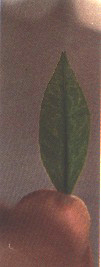
Symptoms. Enations are clearly visible on the underside of leaves as small galls or protuberances on the veins (Figures 98 and 99). When looking for symptoms, it is best to hold the containers so that light is reflected at an angle on the underside of the leaf. Enation symptoms are best seen by reflected light in clear sunlight in the morning or evening hours when the sun is at a lower angle. Enations persist on mature leaves and are generally more prominent on the larger mature leaves.
An additional symptom on the leaves of Mexican lime is leaf cupping, identical to the cupping seen on Mexican lime leaves infected with tristeza (see Figure 6).
Termination. Under conditions of excellent growth flushes, proper temperatures and good nutrient balance, symptoms should be evident within the first three to four flushes of growth or within 12 weeks after inoculation. If positive controls are available, these can be used as a basis of judgement. If not, then train plants as a single vigorous shoot after the second flush and observe new growth for two additional flushes, or until plants reach about 1 m in height. If, during this period, symptoms are not seen, and temperatures in the index facility were cool, the test or index can be terminated.
VEIN-ENATION DETECTION
Summary
Graft transmission
Indicators:
Mexican lime or sour orange seedlings.
No. plants/test:
4 (3 plus 1 control in each of 2 containers).
Inoculum:
"Buds" (buds, blind buds or chip buds).
Plant growth:
Allow all shoots to develop for the first two or three growth
flushes, then train to a single shoot.
Temperature:
Cool: 24-27°C max. day/18-21 °C min. night (or cooler).
First symptoms:
6 to 8 weeks.
Symptoms:
Enations or protuberances on veins on underside of leaves.
REFERENCES
Bazan de Segura, C. & Ferrand, A.1969. Woody gall, its distribution and importance in new and old plantings in Peru. In Proc. 1.st Int. Citrus .Symp.. 3: 1449-1451.
Calavan, E.C., Roistacher, C.N. & Nauer, E.M.1972. Thermotherapy of citrus for inactivation of certain viruses. Plant Dis. Rep., 56: 976-980.
Fraser, L.R.1959. Woody gall, a suspected virus disease of rough lemon and other citrus varieties. In Prac. Linn. Sot. NSW /959, Vol. LXXXIV, 3: 332-334.
Fraser, L.R. & Broadbent, P.1979. Virus and virus-related diseases of citrus in New South Wales. Vein enation-woody gall. In Dept. Agr. NSW publication, p. 50-51. Surrey Beatty & Sons, Chipping Norton, NSW 2170, Australia.
Hooper, G. & Schneider, H.1959. The vein-enation and woody gall disease of citrus. Citrograph, 54: 423-424.
Koizumi, M. & Sasaki, A.1980. Protection phenomena against tristeza in trees preinoculated with vein-enation virus. In Prac. 8th Conf. IOCV, p. 48-50. Riverside, IOCV.
McClean, A.P.D.1954. Citrus vein-enation virus. South Afr . .J. Sci., 50: 147- 151.
Rogers, G.M. & da Graca, J.V.1986. Virus-like particles in citrus vein-enation virus-infected tissue. In Proc. Electron Microsc.. Soc. South. Afi., 16: 127-1213.
Roistacher, C.N. & Calavan, E.C.1974. Inactivation of five citrus viruses in plants held at warm glasshouse temperatures. Plant Dis. Rep., 58: 850-853.
Roistacher, C.N. & Kitto, S.L.1977. Elimination of additional citrus viruses by shoot-tip grafting in vitro Plant Dis. Rep., 61: 594-596.
Wallace, J.M. & Drake, R.J.1953. A virus induced vein enation in citrus. Citrus Leaves, 33(2): 22,24.
Wallace, J.M. & Drake, R.J.1960. Woody gal Is on citrus associated with vein-enation virus infection. Plant Dis. Rep., 44: 580-584.
Wallace, J.M. & Drake, R.J.1961. Induction of woody galls by wounding of citrus infected with vein-enation virus. Plant Dis. Rep., 45: 682-686.
Weathers, L.G. & Greer, F.C., Jr.1967. The use of synergy to demonstrate variants of citrus woody gall virus. Phytopathol., 57: 1388-1389.
Weathers, L.G. & Harjung, M.K.1964. Transmission of citrus viruses by dodder, Cuscuta sublinclusa. Plant Dis. Rep., 48: 102-103.